
Authors: Deepa Srinivasa, Dr. Ramchandra Patil, Bharanidharan K, Prathamesh Bare
Unveiling Rare Phosphorescence in CVD Lab Grown Diamonds: A Gemological Breakthrough
The world of diamonds is undergoing a profound transformation, largely driven by the remarkable advancements in technology that allow for the creation of exquisite gemstones in controlled laboratory environments. Lab Grown Diamonds (LGDs) have surged in popularity, offering a sustainable and often more affordable alternative to their mined counterparts. These technologically advanced diamonds are not mere imitations; they are chemically, physically, and optically identical to natural diamonds, distinguished primarily by their origin and growth kinetics. However, as the sophistication of their production methods evolves, so does the complexity of their characterization, making advanced gemological analysis more crucial than ever for accurate identification and classification.
The Rise of Lab Grown Diamonds and Their Production Methods
Lab Grown Diamonds represent a pinnacle of modern materials science. Produced within days or weeks, these diamonds offer significant investment benefits while maintaining the same dazzling properties. The burgeoning market for LGDs caters to consumers who prioritize transparency, ethical sourcing, and value without compromising on quality or brilliance. Two primary methods dominate the production of these synthetic diamonds: High Pressure High Temperature (HPHT) and Chemical Vapor Deposition (CVD).
The HPHT method mimics the natural conditions deep within the Earth, using extreme pressure (typically around 5.5 GPa) and high temperatures (1300-1600 °C) to crystallize carbon into diamond form. This process often involves a metal solvent/catalyst. In contrast, the CVD method cultivates diamonds from a hydrocarbon gas mixture in a vacuum chamber at much lower pressures and temperatures. Carbon-containing gases, like methane, are broken down into their constituent atoms by microwaves or plasma, allowing carbon atoms to deposit layer by layer onto a diamond seed crystal. Each method leaves behind distinct growth patterns and internal characteristics, making it possible for skilled gemologists and well-equipped laboratories to differentiate them from natural diamonds and from each other, a process vital for market integrity and consumer confidence.
Distinguishing Features and Advanced Gemological Identification
One of the key distinguishing features in CVD lab-grown diamonds often relates to their reaction under ultraviolet (UV) light. When exposed to a conventional Short-Wave Ultra-Violet (SWUV) lamp (280-315 nm), CVD diamonds typically exhibit distinct fluorescence. Common SWUV reactions observed in these synthetic diamonds include orange, yellow, green, violet, or blue fluorescence colors. Prolonged exposure to SWUV can sometimes induce a change in fluorescence color, a phenomenon that offers valuable insights into the diamond’s internal structure and defect centers (Eaton-Magaña & Shigley 2016, Wang et al., 2003, 2005, 2007, 2010, 2012). Understanding these reactions, along with other advanced analytical techniques, is fundamental for accurate diamond identification, origin determination, and classification in modern gemology, especially in discerning post-growth treatments that can alter a diamond’s appearance.
A Unique Case Study: The 1.547 ct CVD Emerald Cut Diamond
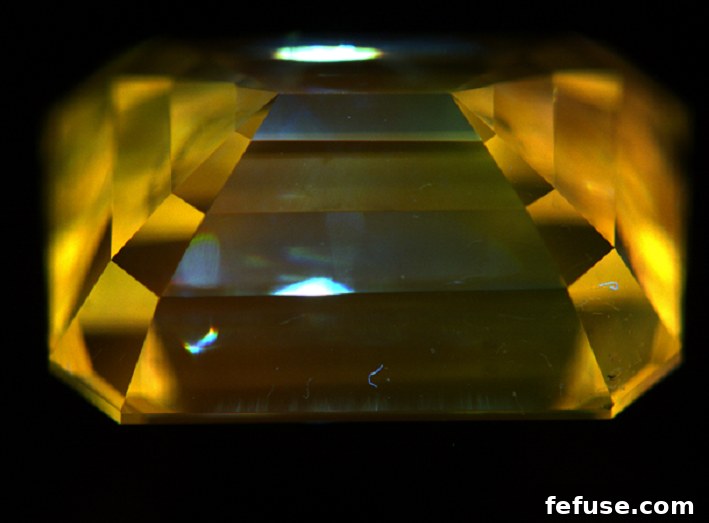
Our recent investigations were captivated by a particular CVD lab-grown diamond weighing 1.547 carats, featuring an elegant emerald cut, VS1 clarity, and an H color grade. This exceptional specimen was submitted to the Gemological Science International (GSI) Mumbai Lab for a critical purpose: Post Growth Treatment Identification. The careful analysis of such diamonds is vital, as post-growth treatments, often involving high-temperature, high-pressure (HPHT) annealing, can modify a diamond’s color and clarity by altering its defect structure. Understanding these treatments is paramount for transparency and accurate valuation in the ever-evolving diamond market, ensuring that consumers and traders are fully aware of a diamond’s complete history.
Advanced Spectroscopic Analysis Unveils Hidden Characteristics
To thoroughly characterize this intriguing diamond, a suite of advanced spectroscopic techniques was employed. Each method provided unique insights into its atomic structure, impurity levels, and the intricate landscape of its defect centers, allowing for a comprehensive understanding of its growth and treatment history.
Mid-Infrared Absorption Spectroscopy (FTIR)
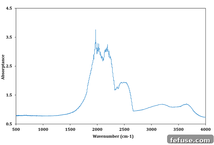
The absorption spectra in the mid-infrared region, meticulously examined using Fourier-transform infrared (FTIR) spectroscopy, provided a foundational understanding of the diamond’s fundamental type. It revealed typical absorption features indicative of a Type IIa diamond. Type IIa diamonds are characterized by the absence of measurable nitrogen impurities, or nitrogen being present only in very low concentrations. This is a common characteristic found in many CVD lab-grown diamonds, as the growth process is meticulously controlled to minimize nitrogen incorporation during deposition. Crucially, no additional anomalous absorption features were observed in this specific sample, suggesting a relatively pure carbon lattice free from other significant impurities that might have arisen from the growth environment or subsequent treatments. This baseline FTIR analysis solidified the diamond’s classification and set the stage for deeper investigations into its defect structures. (See Fig. 1)
Photoluminescence Spectroscopy with 532 nm Excitation
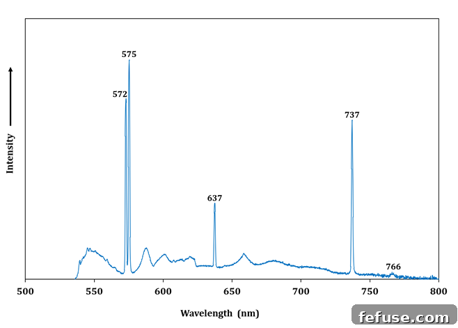
Photoluminescence (PL) spectroscopy, utilizing a 532 nm laser excitation, proved instrumental in identifying specific defect centers within the diamond’s crystal lattice. The resulting spectrum showcased prominent emission peaks at 737 nm, 637 nm, and 575 nm. The emission systems at 737 nm and 766 nm (often appearing as a broader band with the 737 nm peak dominating) are distinctly associated with silicon-vacancy (Si-V) centers. These defects are frequently observed in CVD diamonds, often due to the unintentional etching of silicon from the growth substrate or chamber walls during the high-temperature deposition process. Furthermore, the moderately strong emission lines at 575 nm and 637 nm are characteristic of nitrogen-vacancy (NV) centers, specifically corresponding to the NV0 (neutral) and NV– (negatively charged) states, respectively. The presence and relative intensities of these NV centers are crucial indicators for assessing the growth conditions and potential post-growth annealing treatments of CVD diamonds, as nitrogen is often introduced intentionally or unintentionally during growth and can be mobilized or converted into NV centers during high-temperature post-growth processing (Clark et al., 1995). The specific ratios and intensities of these peaks offer a spectroscopic fingerprint of the diamond’s history. (See Fig. 2)
Raman Microscope Spectroscopy with 532 nm Spectrometers
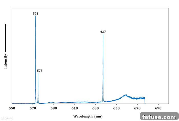
Complementing the PL analysis, Raman microscope spectroscopy, also employing a 532 nm laser, further confirmed the presence of extremely strong NV emission lines at 575 nm and 637 nm. This technique is highly sensitive to the vibrational modes of the crystal lattice and the electronic transitions associated with specific defects. A critical observation from the Raman spectrum was the distinct absence of the 596/597 nm doublet. This doublet, if present, is typically associated with isolated substitutional nitrogen atoms (P1 centers) that have not been converted into more complex nitrogen-vacancy defects. Its absence is a robust and widely recognized indicator of a post-growth treatment, specifically high-temperature annealing. During annealing, P1 centers can diffuse and aggregate, or react with vacancies created during growth to form NV centers, leading to the disappearance of the 596/597 nm signature and a notable enhancement of NV-related emissions. This conclusive spectroscopic evidence unequivocally pointed towards the diamond having undergone significant post-growth treatment, most likely to improve its optical properties or color grade by altering the nitrogen-related defect landscape (Clark et al., 1995). (See Fig. 3)
Revelations from DiamondView Imaging: A Glimpse into Anisotropy and Unique Luminescence
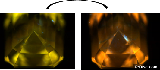
The DiamondView instrument, a specialized short-wave UV imaging system, provided captivating visual insights into the diamond’s internal growth structures and luminescence properties. This powerful tool uses high-energy UV radiation to stimulate fluorescence and phosphorescence, revealing growth patterns that are often invisible under standard magnification. When observed lengthwise, the diamond exhibited a strong greenish-yellow fluorescence, indicating specific defect distributions within those growth sectors. Intriguingly, upon a 180-degree rotation of the diamond, a distinctly strong orange fluorescence was observed. This dramatic and directional change in fluorescence color is indicative of anisotropic growth sectors and the varying distribution of luminescence-activating defects within the crystal lattice, reflecting the unique layer-by-layer growth characteristic of CVD diamonds and their often complex, non-uniform defect incorporation. (See Fig. 4)
The Unprecedented Phosphorescence Discovery: A New Frontier in CVD Diamond Characterization
Typically, most CVD diamonds examined under DiamondView display fluorescence colors such as orange, red, blue, or sometimes a mottled distribution of purple, red, and blue, reflecting the diversity of their defect centers. Phosphorescence, the emission of light that continues after the excitation source is removed, is usually inert or very weak in CVD diamonds, often appearing as a faint blue-green in rare instances. These variations in fluorescence color and phosphorescence intensities are generally understood to be related to the specific growth parameters and defect concentrations within the CVD growth layers, as well as any subsequent treatments (Lu, Q. et al., 2021). The internal crystal plane defects are believed to play a significant role in influencing these observable luminescence characteristics.
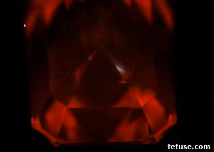
However, during our prolonged observation of the 1.547 ct CVD diamond in the DiamondView, an entirely unprecedented phenomenon was observed. Immediately after the DiamondView’s UV excitation was turned off, an intense red phosphorescence became strikingly visible for a brief period – approximately 10-15 milliseconds – before rapidly transitioning to a distinct yellow glow. This striking and vivid red phosphorescence, particularly its intensity, transient nature, and subsequent color shift, has not been previously reported in CVD diamonds in scientific literature or extensive gemological observations. It was noted that this intense red phosphorescence only manifested after extended exposure to the DiamondView’s high-energy UV light, suggesting a dynamic interaction between the UV radiation and specific, perhaps metastable, defect centers within the diamond. This observation opens new avenues for understanding the complex physics of defects in synthetic diamonds. (See Fig. 5)
Discussion and Implications of the Findings: Unraveling Defect Chemistry
The multifaceted luminescence behavior observed in this CVD diamond – the directional changes in fluorescence and, most notably, the rare intense red phosphorescence – strongly suggests the presence of highly complex and inconsistently distributed lattice defects. These defects likely originate during the intricate CVD growth process, potentially exacerbated or modified by subsequent post-growth treatments like annealing. The varying fluorescence colors observed along different directions indicate significant anisotropy within the crystal, where defect concentrations or configurations differ across distinct growth sectors. This anisotropy influences how the diamond absorbs and re-emits light, leading to the observed color shifts with rotation, providing a visual signature of the diamond’s layered growth.
The discovery of the intense, short-lived red phosphorescence is particularly significant for gemological science. While phosphorescence in diamonds is generally understood to arise from the delayed recombination of electrons and holes trapped at various defect sites, the specific mechanism for this vibrant red emission and its rapid transition to yellow remains an intriguing subject for further research. It is plausible that prolonged UV exposure leads to the population of deep trap states, which then release energy over milliseconds, producing the red glow. The subsequent yellow phosphorescence might arise from shallower traps or different defect complexes, or a de-excitation pathway through another defect center. This observation not only adds a new, distinctive characteristic to the identification toolkit for CVD diamonds but also deepens our understanding of the complex interplay between growth parameters, impurity incorporation, defect chemistry, and optical phenomena in synthetic diamond materials. Such unique features could potentially serve as novel markers for specific growth conditions, post-growth treatment regimes, or even the origin of the CVD diamond, providing an even finer resolution in gemological differentiation and helping to combat undisclosed treatments.
Conclusion: Advancing Gemological Characterization and Scientific Understanding
This comprehensive gemological study of a 1.547 ct emerald cut CVD lab-grown diamond, combining detailed FTIR, Photoluminescence, Raman, and DiamondView analyses, has yielded critical insights into its post-growth treatment and, more profoundly, uncovered a previously unrecorded luminescence phenomenon. The spectroscopic data conclusively confirmed the diamond’s Type IIa nature and the application of post-growth treatment, evidenced by the characteristic presence of NV and Si-V centers and the notable absence of the 596/597 nm doublet. However, the most compelling and scientifically significant finding was the observation of an intense, transient red phosphorescence that rapidly shifted to yellow, visible for milliseconds after prolonged UV excitation in the DiamondView. This unprecedented characteristic highlights the evolving complexity of lab-grown diamonds and the continuous need for advanced gemological research to keep pace with technological innovations in their production.
The unique optical behavior observed in this diamond underscores the intricate relationship between growth defects, impurity profiles, and luminescence properties in CVD materials. It strongly suggests that specific, possibly metastable, defect structures are activated by prolonged high-energy UV exposure, leading to this distinct and dynamic phosphorescence signature. This discovery not only enriches our fundamental understanding of CVD diamond physics but also provides a potential new indicator for distinguishing various growth conditions or post-growth treatments, thereby enhancing the precision of diamond characterization. As lab-grown diamonds continue to gain market share and their production methods become increasingly sophisticated, such detailed scientific investigations are indispensable for maintaining consumer confidence, ensuring accurate identification, and continuously pushing the boundaries of gemological science and diamond research.
References
Wang W., Moses T., Linares R., Shigley J.E., Hall M., Butler J.E. (2003) Gem-quality synthetic diamonds grown by a chemical vapor deposition (CVD) method. G&G, Vol. 39, No. 4, pp. 268– 283, http://dx.doi.org/10.5741/GEMS.39.4.268
Wang W., Smith C.P., Hall M.S., Breeding C.M., Moses T.M. (2005) Treated-color pink-to-red diamonds from Lucent Diamonds Inc. G&G, Vol. 41, No. 1, pp. 6–19, http://dx.doi.org/10.5741/GEMS.41.1.6
Wang W., Hall M.S., Moe K.S., Tower J., Moses T.M. (2007) Latest generation CVD-grown synthetic diamonds from Apollo Diamond Inc. G&G, Vol. 43, No. 4, pp. 294–312, http://dx.doi.org/10.5741/GEMS.43.4.294
Wang W., Doering P., Tower J., Lu R., Eaton-Magaña S., Johnson P., Emerson E., Moses T.M. (2010) Strongly colored pink CVD lab-grown diamonds. G&G, Vol. 46, No. 1, pp. 4–17, http://dx.doi.org/10.5741/GEMS.46.1.4
Wang W., D’Haenens-Johansson U.F.S., Johnson P., Moe K.S., Emerson E., Newton M.E., Moses T.M. (2012) CVD synthetic diamonds from Gemesis Corp. G&G, Vol. 48, No. 2, pp. 80– 97, http://dx.doi.org/10.5741/GEMS.48.2.80
Eaton-Magaña, S. & Shigley, J.E. 2016. Observations on CVD-grown synthetic diamonds: A review. Gems & Gemology, 52(3), 222–245, https://doi.org/10.5741/gems.52.3.222.
Clark, C.D., Kanda, H., Kiflawi, I., and Sittas, G., 1995. Silicon defects in diamond. Physical Review B, 51, 16681–8.
Lu, Q.; Gong, H.; Guo, Q.; Huang, X.; Cai, J. Gemological Characteristic Difference between Colorless CVD Synthetic Diamonds and Natural Diamonds. Materials 2021, 14, 6225. https://doi.org/10.3390/ma14206225.