
Revolutionizing Disease Detection: Graphene-Gold Biosensors Achieve Near-Perfect Efficiency
The dawn of a new era in medical diagnostics is upon us, heralded by a groundbreaking innovation that combines the extraordinary properties of graphene and gold. Scientists have engineered ultrasensitive biosensors capable of detecting diseases at the molecular level with near-perfect efficiency, promising to transform how we diagnose and combat some of the most challenging health conditions facing humanity and the animal kingdom.
This remarkable advancement, spearheaded by researchers at the University of Minnesota, represents a significant leap forward in biosensor technology. Their work, detailed in a seminal paper published in the esteemed journal Nature Nanotechnology, describes the development of devices that can meticulously probe protein structures. This capability is crucial, as it allows for the early and accurate detection of a wide array of disorders directly linked to protein misfolding.
The implications of this research are vast, extending to devastating conditions such as Alzheimer’s disease in humans, as well as chronic wasting disease and mad cow disease prevalent in animals. These protein-misfolding disorders pose immense diagnostic and therapeutic challenges, and the development of highly sensitive, reliable detection methods is paramount for effective management and potential cures.
The Critical Need for Ultrasensitive Disease Detection
In the realm of medical science, early detection is often the most powerful weapon against disease. For many conditions, particularly those involving subtle molecular changes, the ability to identify biomarkers at extremely low concentrations can make the difference between successful intervention and irreversible progression. Biosensors are at the forefront of this battle, serving as sophisticated tools designed to detect specific biological molecules or events. However, conventional biosensors often struggle with the level of sensitivity required for the earliest stages of complex diseases, especially when dealing with the intricate world of protein structures.
Protein misfolding, a process where proteins adopt an abnormal shape, is a fundamental mechanism underlying a diverse group of debilitating neurodegenerative and systemic diseases. Detecting these misfolded proteins early and understanding their structural nuances is key to unraveling disease pathogenesis and developing targeted therapies. Unfortunately, current methodologies face numerous technical hurdles in achieving the necessary precision and sensitivity to probe these minute, yet critical, molecular alterations. The quest for more accurate and earlier detection methods has driven extensive research into novel materials and advanced fabrication techniques.
Graphene: A Material of Immense Promise and Intrinsic Challenge
Graphene, a single layer of carbon atoms arranged in a hexagonal lattice, has long been hailed as a wonder material due to its extraordinary properties. Known for its exceptional strength, conductivity, and atomic thinness, graphene has garnered significant attention across various scientific fields, including biosensing. Its unique two-dimensional structure makes it an ideal candidate for interacting with biological molecules at the nanoscale, offering a high surface-to-volume ratio crucial for sensitive detection.
Indeed, graphene has already found applications in biosensors, demonstrating its potential to detect various biomolecules. However, its very strength—its single-atom thickness—also presented a significant paradox and a major hurdle for optical biosensing. As Sang-Hyun Oh, the lead researcher on the study, explained in a media statement, “In order to detect and treat many diseases we need to detect protein molecules at very small amounts and understand their structure. Currently, there are many technical challenges with that process. We hope that our device using graphene and a unique manufacturing process will provide the fundamental research that can help overcome those challenges.”
The primary issue stemmed from graphene’s remarkably thin nature: it does not interact efficiently with light. For sensitive molecular detection, efficient light absorption and subsequent conversion into local electric fields are absolutely essential. Previous research utilizing graphene nanostructures, despite their innovative design, could only achieve a light absorption rate of less than 10%. This inherent inefficiency limited graphene’s full potential in optical biosensing, prompting scientists to seek novel ways to enhance its light interaction capabilities without compromising its other advantages.
The University of Minnesota’s Groundbreaking Synergy: Graphene and Gold
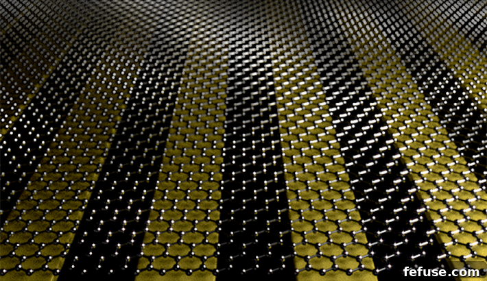
Recognizing the limitations of graphene alone, the University of Minnesota researchers embarked on an innovative path to amplify its performance. Their breakthrough involved a synergistic approach: combining the unparalleled properties of graphene with nano-sized metal ribbons of gold. This ingenious pairing capitalized on the strengths of both materials while cleverly mitigating graphene’s optical shortcomings.
The team’s method was as elegant as it was effective. To create an ideal platform for their novel biosensors, they employed a high-tech nanofabrication technique known as “template stripping,” coupled with the surprisingly simple use of sticky tape. This combination allowed them to construct an ultra-flat base layer surface for the graphene. The precision and flatness achieved through this method were critical, as even minute imperfections at the nanoscale can significantly impact the efficiency and reliability of biosensors designed for molecular detection.
Engineering Precision: The “Template Stripping” Technique
The “template stripping” technique is a sophisticated nanofabrication process that is fundamental to the UMN team’s success. It involves creating extremely smooth and pristine metal surfaces, typically gold or silver, by stripping them from a highly polished master template. This method minimizes surface roughness to an unprecedented degree, creating an atomically flat substrate. When combined with graphene, this ultra-flat gold surface provides a stable and consistent environment that is crucial for subsequent optical and molecular interactions. The flatness ensures that the delicate molecular structures being probed are not distorted by an uneven substrate, thus enabling higher resolution and more accurate detection.
The meticulous engineering of this base layer was a pivotal step, setting the stage for the dramatic improvement in light absorption and interaction that followed. The integration of gold nano-ribbons provided a platform that not only enhanced the mechanical stability of the graphene but also introduced plasmonic properties crucial for overcoming the optical limitations of graphene.
Unlocking Unprecedented Efficiency: Harnessing Plasmon Generation
With their meticulously prepared graphene-gold platform, the researchers then focused on enhancing the interaction between light and the single-atom-thick graphene layer. Their ingenious strategy involved using the energy of light to generate a phenomenon known as plasmons—collective oscillations of electrons, often described as “sloshing motions” within the material. This technique, when applied to their novel device, yielded astonishing results.
By shining light onto the graphene-gold composite, the scientists were able to create a plasmon wave with unprecedented efficiency. This wave resulted in a near-perfect 94 percent light absorption, transforming the incident light into what the university’s press release vividly described as “tidal waves” of an electric field. This level of light absorption is a monumental leap from the less than 10% previously achieved with graphene alone, marking a paradigm shift in the material’s utility for optical sensing.
The implications of such high light absorption were immediately apparent. When protein molecules were carefully inserted between the graphene and metal ribbons, the device was able to harness enough energy from these amplified electric fields to enable the direct viewing of single layers of protein molecules. This capability is extraordinarily significant, as it moves beyond simply detecting the presence of a molecule to providing insights into its structural arrangement at a resolution previously unattainable.
The scientific community, including the UMN team itself, was particularly struck by the accuracy and consistency of their findings. According to Oh, he and his team were genuinely surprised by the rate of light absorption, which matched their sophisticated computer simulations almost perfectly. This strong correlation between experimental results and theoretical predictions provides robust validation for their innovative design and the underlying physical principles.
A New Era for Diagnosing Protein Misfolding Disorders
The enhanced capabilities of these graphene-gold biosensors herald a new era for the diagnosis and study of protein misfolding disorders. Diseases like Alzheimer’s, Parkinson’s, and Creutzfeldt-Jakob disease in humans, along with conditions like chronic wasting disease (CWD) and bovine spongiform encephalopathy (BSE, or mad cow disease) in animals, are all characterized by the accumulation of misfolded proteins. These misfolded proteins, often referred to as prions or amyloid aggregates, are notoriously difficult to detect in their earliest, most crucial stages due to their low concentrations and complex structures.
Current diagnostic methods for these disorders often rely on indirect markers, late-stage symptoms, or invasive procedures, which can delay diagnosis and treatment. The ability of the new biosensors to directly probe and visualize single layers of protein molecules offers a superior solution. This technology could enable the detection of misfolded proteins far earlier than previously possible, potentially years before clinical symptoms manifest. Such early detection is critical for initiating timely interventions, which could involve new therapeutic strategies aimed at preventing further protein aggregation or clearing existing misfolded proteins.
Furthermore, the device’s capacity to understand protein structure at a molecular level opens avenues for fundamental research into the mechanisms of these diseases. By observing how proteins misfold and aggregate in real-time and at high resolution, scientists can gain invaluable insights into disease progression, identify potential therapeutic targets, and develop more effective drugs. This could dramatically accelerate drug discovery pipelines for currently incurable conditions, offering hope to millions affected by these devastating illnesses.
The Broader Implications and Future of Nanosensors
While the immediate application of these graphene-gold biosensors lies in diagnosing protein misfolding disorders, their broader implications extend far beyond this specific area. The fundamental principles demonstrated by this research—achieving ultra-high sensitivity and efficient light-matter interaction at the nanoscale—could revolutionize various fields that rely on molecular detection.
In personalized medicine, these advanced biosensors could enable comprehensive biomarker profiling, allowing for highly individualized disease risk assessment and treatment strategies. For drug discovery, they could accelerate the screening of new compounds by providing rapid and accurate assessments of drug-protein interactions. Environmental monitoring could benefit from ultrasensitive detection of pollutants at trace levels, while food safety could be enhanced through rapid pathogen and contaminant identification.
The journey from a laboratory breakthrough to widespread clinical application is often long and complex. However, the scientists are highly optimistic that this technique will significantly improve various devices used to detect disorders related to protein misfolding. Future work will likely focus on miniaturization of the devices, optimizing the manufacturing process for scalability and cost-effectiveness, and conducting extensive validation studies in clinical settings. The potential for these advanced nanosensors to be integrated into point-of-care diagnostics, allowing for rapid and accurate testing outside traditional laboratory environments, is particularly exciting.
Conclusion: A Glimmer of Hope for Molecular Diagnostics
The pioneering research from the University of Minnesota marks a pivotal moment in the advancement of biosensor technology. By ingeniously combining graphene and gold, scientists have overcome long-standing challenges in optical biosensing, achieving an unprecedented 94% light absorption efficiency. This breakthrough has not only enabled the visualization of single protein layers but also paved the way for the ultrasensitive detection of complex protein misfolding disorders.
This innovative technology offers a powerful new tool in the fight against devastating diseases like Alzheimer’s and chronic wasting disease, promising earlier diagnosis, deeper mechanistic understanding, and ultimately, more effective treatments. As this fundamental research continues to evolve, the transformative potential of graphene-gold biosensors stands as a beacon of hope, set to redefine the landscape of molecular diagnostics and significantly improve both human and animal health in the years to come.